


| Address |
| Department of Biointerface Chemistry, Faculty of Pharmaceutical Sciences, University of Toyama 2630 Sugitani, Toyama, 930-0194, Japan Phone: +81-76-434-7565 |


Biological membranes play the role as the interface that separates the inside and outside of cells and organelles, and as the sphere of various vital activities such as energy conversion, material transport, and information transmission. Living organisms have acquired a boundary membrane that distinguishes themselves from the outside world by self-organization of lipid molecules, being endowed with physical flexibility and substance-selective permeability that are impossible with rigid walls. The fact that thousands of lipid species are present in the body clearly indicates that the unique biochemical properties of each lipid provide a high level of functions to the body. However, taking the fact that biomembranes are exactly where these functions are exerted into account, we should note that all life activities are supported by the physicochemical properties of biological membranes as molecular assemblies.
Based on this perspective, we are conducting researches from the aspects of the biophysics and biointerface chemistry, to clarify the biological phenomena on biological membranes both in terms of spatial structure (of angstroms to micrometers) and temporal structure (of nanoseconds to minutes). Phospholipids, the main components of biological membranes, spontaneously assemble to form lipid bilayers. These membranes form a dynamic and heterogeneous structure in which the molecules fluctuate due to thermal motion. In addition, molecular metabolism and transport also modify the composition and molecular localization of membranes all the time. Proteins, another important component of biological membranes, are deeply involved in this metabolism and transport. We aim to measure the complex spatiotemporal structure of the biological interface with lipids and proteins and to explain how the biological membranes play a vital role in terms of physical chemistry and colloidal science. In addition to basic scientific aspects related to the elucidation of biological phenomena of membranes, we are also conducting researches aimed at application for the pharmaceutical preparations through the analysis of microparticles of lipid-protein complexes.
1. Interbilayer transfer dynamics of lipids
Most of the lipids in the body are synthesized in the endoplasmic reticulum and distributed to the plasma membrane and various organelle membranes. Lipids secreted from the liver and small intestine circulate in the blood as lipoproteins and are supplied to peripheral cells. Thus, intracellular and intercellular lipid transport plays an important role in maintaining homeostasis. However, quantitative discussions on lipid dynamics have not been carried out well not only in cell systems but also in cell-free systems. This is because no rational dynamics measurement methods were available. We use phospholipid vesicles (liposomes) to kinetically analyze the intervesicular lipid transfer, using fluorescence resonance energy transfer (FRET), intermolecular excimer fluorescence, and neutron scattering. Currently, we are trying to apply them as assay systems for proteins with lipid transfer activity.
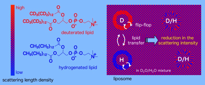
Evaluation of phospholipid transfer dynamics by neutron scattering.
2. Flip-flop of phospholipids
The lipid composition of the plasma membrane is greatly different between the cytoplasmic side and the outer side of the bilayer. In apoptosis, the asymmetry is broken and the phosphatidylserine localized on the cytoplasmic leaflet is exposed to the outside. In apoptosis, the asymmetry is broken, and the phosphatidylserine localized on the cytoplasm leaflet is exposed to the extracellular leaflet, which becomes a signal for phagocytosis by macrophages. In other words, disruption of the plasma membrane asymmetry is directly linked to cell death. In the endoplasmic reticulum, on the other hand, newly synthesized phospholipids are introduced into the cytoplasmic side of the lipid bilayer, and then rapidly transferred (fliped) to the luminal leaflet to maintain the balance of the number of lipids between both leaflets. In this way, control of lipid dynamics is an essential issue in life activities. We are conducting research to evaluate the rate of the phospholipid flip-flop using fluorescence quenching and neutron scattering, and to clarify the factors involved in the promotion of the flip-flop. We are also constructing a system that artificially produces asymmetry to the liposome membranes.

Symmetric and asymmetric distribution of phospholipids in biomembranes.
3. Nanodisc
High-density lipoprotein (HDL) is a colloidal particle in the blood responsible for the reverse cholesterol transport system, which transports peripheral cholesterol to the liver. HDLs are produced by the liver and peripheral cells and become disc-shaped initially when produced. These discoidal particles can be also created artificially (which are called nanodiscs) using phospholipids and apolipoprotein A-I (apoA-I), which is a major protein component in HDL, or amphiphilic polypeptides having a similar structure to apoA-I. We are elucidating the structure of nanodiscs using various fluorescence analysis methods. By the fluorescence lifetime and intramolecular excimer fluorescence, we have found that small nanodiscs with low phospholipid content have a lipid bilayer that is not planar but forms a saddle-shaped curved surface. Neutron scattering analysis revealed that the phospholipid dynamics are significantly different between nanodiscs and vesicles and that the dissociation rate of phospholipids and cholesterol from nanodiscs is significantly faster than that from vesicles. In addition, we are constructing nanodisc systems in which nanodiscs are self-replicated or stabilized by peptide ligation. Nanodiscs are not only physiologically important but are also being applied to membrane protein remodeling tools and drug delivery systems. Clarifying the physicochemical properties of nanodiscs is, therefore, very significant.
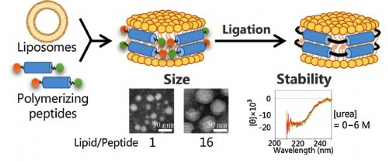
Nanodisc with higher stability acquired by the peptide ligation (Colloid Surf. B 146, 423 (2016)).
4. Membrane curvature and protein-membrane interaction
Biological membranes greatly change the curvature of the membrane during processes such as membrane fusion. Many proteins have been found that induce or recognize the curvature of the membrane. With vesicles with different sizes (curvatures), we are evaluating how curvature changes the membrane environment and how it affects the interaction with proteins. It was clarified by the isothermal titration calorimetry that the enthalpy of the membrane increases with the increase in the curvature. We also found that the binding of amyloid β protein to the membrane and amyloid fibrillation are promoted in high curvature membranes. We are conducting research aimed at elucidating the physiological significance of changes in the membrane curvature.
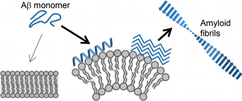
High membrane curvature enhances binding and fibrillation of amyloid β (Langmuir 31, 11549 (2015)).
5. Non-lamellar phase-forming lipids and novel lipid particulates
Biological membranes form a bilayer structure, but they contain several lipids with negative spontaneous curvature called nonlamellar phase-forming lipids. Such lipids are thought to change the lateral pressure in the membrane and regulate the membrane binding of peripheral membrane proteins and the structure and activity of the integral membrane proteins. We have devised a method to detect changes in the lateral pressure of the membrane by fluorescence and NMR, and have clarified the effect of nonlamellar phase-forming lipids on the lateral pressure. Meanwhile, the non-lamellar phases (inverted hexagonal phase and cubic phase) formed by these lipids have been considered difficult to disperse stably in water, but we have reported that their colloidal particles with a 100-200-nm diameter (cubosomes and hexosomes) can be obtained by using polymer emulsifiers. Recently, it has also been found that the liquid crystal structure and interaction with plasma proteins can be controlled depending on lipids and emulsifiers. Elucidation of the physical properties and biophysical functions of the inverted hexagonal phase and cubic phase is progressing, and the way to its pharmaceutical application is also being opened.
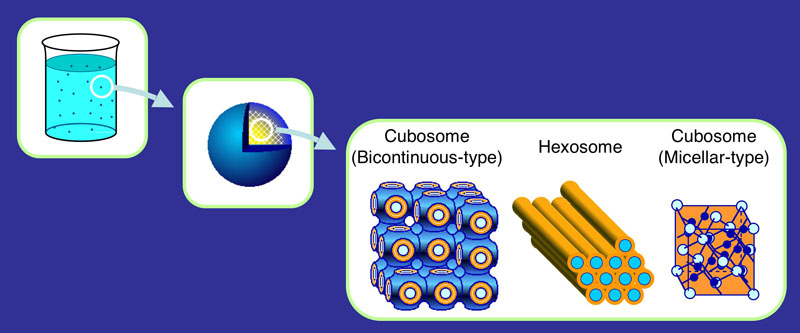
Schematic representation of
cubosome and hexosome